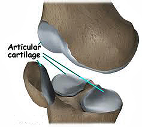
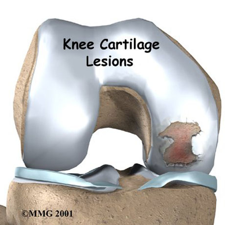
Articular cartilage, also called hyaline cartilage, is the smooth, glistening white tissue that covers the surface of all the diarthrodial joints in the human body. As its name implies, articular cartilage is critical in the movement of one bone against another. Articular cartilage has an incredibly low coefficient of friction which, coupled with its ability to bear very large compressive loads, makes it ideally suited for placement in joints, such as the knee and hip.
Cartilage is composed of specialized cells called chondrocytes that produce a large amount of extracellular matrix composed of Type II collagen (except Fibrocartilage which also contains type I collagen) fibers, abundant ground substance rich in proteoglycan, and elastin fibers. Cartilage is classified in three types, elastic cartilage, hyaline cartilage and fibrocartilage, which differ in the relative amounts of these three main components.
Unlike other connective tissues, cartilage does not contain blood vessels. The chondrocytes are supplied by diffusion, helped by the pumping action generated by compression of the articular cartilage or flexion of the elastic cartilage. Thus, compared to other connective tissues, cartilage grows and repairs more slowly.
ARTICULAR CARTILAGE DAMAGE
Articular cartilage lesions in people and athletic population commonly occur and result from the significant acute and chronic joint stress associated with high impact activities. These lesions have poor intrinsic healing capacity, and the persistent defect in the joint surfaces cause pain, swelling and mechanical symptoms that result in functional impairment and limitation of athletic participation. If untreated, articular cartilage lesions can lead to chronic joint degeneration and disability. Several techniques for articular cartilage repair have been recently developed with promising results. SYMPTOMS
• Patients with focal chondral defects generally have symptoms localized to the lesion location.
• Symptoms that worsen with weight-bearing activities likely are produced by damage that resides in the femoral condyle.
• Pain when arising from a sitting position following prolonged knee flexion and with stair climbing suggests a lesion in the patellofemoral joint.
• Patients may complain of activity-related swelling.
An effusion may be palpable, and the affected joint may feel warmer in the presence of an underlying inflammatory process. The ranges of active and passive motion of the knee without effusion are usually within normal limits in the early stages of articular cartilage disease. Localized joint-line pain may be present on palpation, and the McMurray test may elicit some discomfort in the involved compartment. Associated degenerative meniscal tears and articular cartilage disease are commonly associated with older patients. The knee is usually stable to ligament evaluation in most patients, but excluding underlying ligamentous instability is an important part of the examination and future management. RADIOGRAPHIC IMAGINE
Radiographic imaging of knees with chondral disease should include weight-bearing views for better estimation of mechanical and anatomical axis of lower limb. Magnetic resonance imaging (MRI) is a useful noninvasive means that may help demonstrate symptomatic articular cartilage lesions. There is often a high correlation with radiographs, but MRI may be particularly helpful when standard radiographs appear normal. Current techniques are more accurate in detecting partial or full-thickness cartilage loss and how large is the damage. These information can define the kind of operative technique which is the right for the patient.
Patients with acute chondral injuries are treated as soon as practical after the diagnosis is made, especially if the knee is being treated concurrently for meniscus or anterior cruciate ligament (ACL) pathology.
Patients with chronic or degenerative chondral lesions often are treated nonoperatively (conservatively) for at least 12 weeks after a suspected chondral lesion is diagnosed clinically. This treatment regimen includes activity modification, physical therapy, nonsteroidal anti-inflammatory drugs, viscosupplement injections, and perhaps dietary supplements that may have cartilage-stimulating properties. If nonoperative treatment is not successful, then surgical treatment is considered.
OPERATIVE TREATMENT
Therapy
REHABILITATION
SURGICAL TREATMENT
Many new techniques for the treatment of articular cartilage damage exist with very good results.
MICROFRACTURE TECHNIQUE + AMIC TECHNIQUE
Microfracture technique in Osteochondritis Dissecans
Microfracture techniques combine with collagen substitute Cartilage lesion in the center and low part of the patella Abrasion of the cartilage lesion until the subchondral bone Abnormal cartilage removal
Collagen menbrane insertion in the articular cartilage lesion Collegen membrane with autologous stem cell just before implantation to articular damage.
Autologous stem cells and fibrin glue insertion in the crater of the lesion Articular cartilage lesion grade VI (Outerbrigde) in woman 59 years old. Pre (red circle) and two years postoperative with operative treatment with autologous bone marrow stem cells and collagen membrane (AMIC technique)
Red arrow – cartilage lesion, green arrow two years postop
The same patient.
Microfracture is indicated as first-line treatment for focal Outerbridge grade III and IV lesions under 3–4 cm2 in an otherwise nonarthritic knee in a symptomatic, active, and relatively young patient willing to respect the postoperative rehabilitation requirements.
For older and low-demand patients, microfracture may be useful even in lesions larger than 3–4 cm2
POSTOPERATIVE
Cold therapy is typically used for 1 to 7 days postoperatively.
Crutch-assisted touchdown weight-bearing ambulation (10% of body weight initially) is prescribed for 6 to 8 weeks, depending on the size of the lesion.
For most patients, 6 to 8 weeks is adequate time to limit weight bearing. However, for patients with small lesions (<1 cm diameter), weight bearing may be initiated earlier.
Patients with lesions on the femoral condyles or tibial plateaus rarely use a brace during the initial postoperative period. However, we may prescribe an unloading type brace when the patient becomes more active and the postoperative swelling has resolved.
OSTEOCHONDRAL ALLOGRAFT TRANSPLANTATION
Osteochondral allografts have been successfully used for the treatment of large and deep chondral and osteochondral lesion from acute trauma, osteochondritis dissecans and osteonecrosis.
Better outcomes have been reported in patients with unipolar lesions, without malalignment, rigid fixation and age younger than 50 years. Better outcomes are typically seen in young, active adults as well.
Lesion diameter typically ranges from 15 to 35 mm.
Controversies exist in cases:
• Patients with inflammatory arthritis
• Morbid obesity
• Medical problems that may interfere with the incorporation of the allograft into the host tissue.
POSTOPERATIVE
The early postoperative management include the use of continuous passive motion while the patient is in the hospital.
Patient generally are allowed a full range of motion unless they had undergone additional reconstructive procedures such a meniscal repair, anterior cruciate ligament reconstruction or osteotomy that would alter the rehabilitation plan.
Patients are allowed touch-down weight bearing for a minimum of 6 to 8 weeks postoperatively, or until bony union is determined by radiographs.
For femoral condyle allografts, no bracing is needed; but if an osteotomy is performed, a hinged range-of-motion brace is used for protection until healing is apparent. Weight bearing is progressed slowly between the second and fourth month, with full weight bearing using a cane or crutch.
AUTOLOGOUS CHONDROCYTE IMPLANTATION – STEM CELLS
Injuries to joint surfaces can result from acute high-impact or repetitive shear and torsional loads to the superficial zone of the articular cartilage architecture. The use of autologous chondrocyte implantation is promising and is associated with several potential long-term benefit.
Autologous chondrocyte implantation is ideally suited for symptomatic deep chondral lesion along the femoral condyle or trochlear region. High-demand patient between 15-55 years of age with excellent motivation and potential for compliance are the best candidates.
However autologous chondrocyte implantation is a viable option for a symptomatic patient with a lesion of >2 sq.cm but <12 sq.cm. and for a patient who continues to have pain after mosaicplasty or microfracture procedure.
Bone involvement is not contraindication, but staged or concomitant autologous bone grafting should be undertaken when the bone involvement is deeper than 6-8 mm.
Autologous chondrocyte transplantation is not a treatment for osteoarthritis (general joint disease), gout, and rheumatoid arthritis or other systemic joint diseases.
Autologous chondrocyte are arthroscopicaly harvested from a less weight-bearing area, commercially extracted from the harvested cartilage (200-500mg), and multiplied in vitro (labor cell culture).
Elective reinplantation is performed 4-6 weeks after cartilage harnesting by debridement of the defect to an intact margin, carelully avoiding osseous bleeding from the bed of the defect.
The lesion area is already clear and ready to receveive the autologous chondrocyte.
Chondrocyte culture.
The surgeon cut the chondrocyte film like the shape of the chondral defect.
The autologous chondrocyte is ready to implant.
The autologous chondrocyte is transplanted. The chondral defect is covered.
Chondral defect due to trauma. The autologous chondlal cell filling the defect.
POSTOPERATIVE
Postoperatively protected weight-bearing are restricted for 6 weeks. The crutches is necessary for walking.
The physical therapy program started 2nd post-op day with passive movement (physical therapist or CPM machine) until 4th week.
Therapy concentrates on quadriceps activation and isometric straight leg raises. After the 6th week the patient can walk without aid devices with full weight-bearing. Full weight bearing and full range of motion are achieved. Closed-chain exercises and functional training are started.
After 12th week Strengthening and functional training are progressed. Return to full activities is delayed for at least 8 months to protect the lesion as it matures.
Return to participation in pivoting sports is usually allowed by 12 months.
MICROFRACTURE TECHNIQUE
Cold therapy is typically used for 1 to 7 days postoperatively.
Crutch-assisted touchdown weight-bearing ambulation (10% of body weight initially) is prescribed for 6 to 8 weeks, depending on the size of the lesion.
For most patients, 6 to 8 weeks is adequate time to limit weight bearing. However, for patients with small lesions (<1 cm diameter), weight bearing may be initiated earlier.
Patients with lesions on the femoral condyles or tibial plateaus rarely use a brace during the initial postoperative period. However, we may prescribe an unloading type brace when the patient becomes more active and the postoperative swelling has resolved.
OSTEOCHONDRAL ALLOGRAFT TRANSPLANTATION
The early postoperative management include the use of continuous passive motion while the patient is in the hospital.
Patient generally are allowed a full range of motion unless they had undergone additional reconstructive procedures such a meniscal repair, anterior cruciate ligament reconstruction or osteotomy that would alter the rehabilitation plan.
Patients are allowed touch-down weight bearing for a minimum of 6 to 8 weeks postoperatively, or until bony union is determined by radiographs.
For femoral condyle allografts, no bracing is needed; but if an osteotomy is performed, a hinged range-of-motion brace is used for protection until healing is apparent. Weight bearing is progressed slowly between the second and fourth month, with full weight bearing using a cane or crutch.
ΑUTOLOGOUS CHONDROCYTE IMPLANTATION
Postoperatively protected weight-bearing are restricted for 6 weeks. The crutches is necessary for walking.
The physical therapy program started 2nd post-op day with passive movement (physical therapist or CPM machine) until 4th week.
Therapy concentrates on quadriceps activation and isometric straight leg raises. After the 6th week the patient can walk without aid devices with full weight-bearing. Full weight bearing and full range of motion are achieved. Closed-chain exercises and functional training are started.
After 12th week Strengthening and functional training are progressed. Return to full activities is delayed for at least 8 months to protect the lesion as it matures.
Return to participation in pivoting sports is usually allowed by 12 months.