ACUTE PATELLAR DISLOCATION

TRAUMATIC PATELLAR DISLOCATION

Patellar dislocation occur as a result of a twisting injury with strong contraction of the quadriceps. 
An acute effusion may develop, and this diagnosis may be mistaken for the more common anterior cruciate ligament injury. The patella usually reduced easily with extension of the knee, and the patient may never have appreciated that the patella was dislocated.

Acute injury often are accompanied by a large effusion and knee tenderness. Occasionally the patella may be so unstable that simple range of motion may displace it.


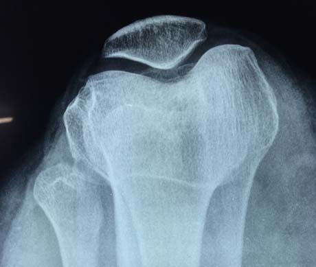
X-Ray of the right knee.  The normal position of the patella in the knee joint

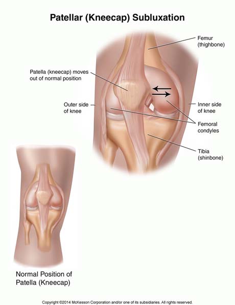
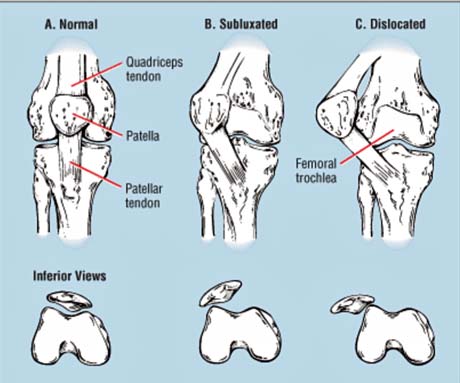
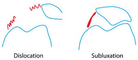
Up: Patellar lateral dislocation
Down: Normal patellar position.


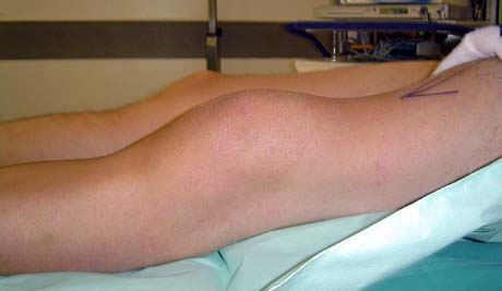
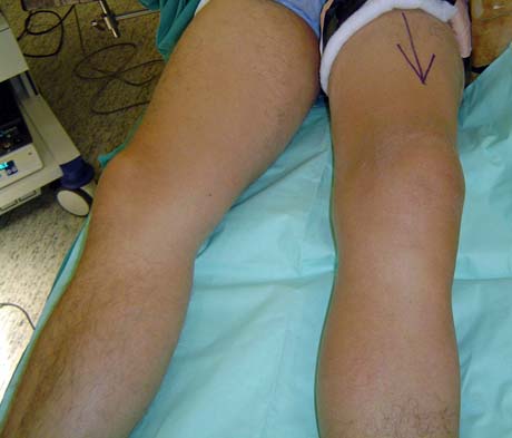
The injured left knee is swollen due to patellar dislocation.

The medial structures is ruptured after acute patellar dislocation.

The patella is dislocated laterally.  The medial patelofemoral ligament was torn.

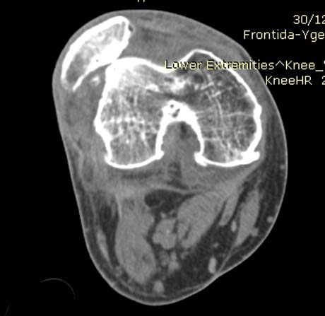
Right knee MRI.  Lateral patellar dislocation due to trauma


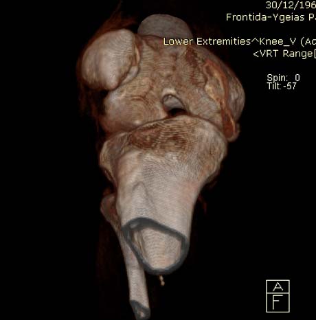
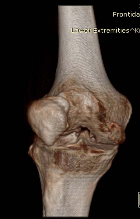
3D reconstruction of the right knee.  Traumatic patellar dislocation

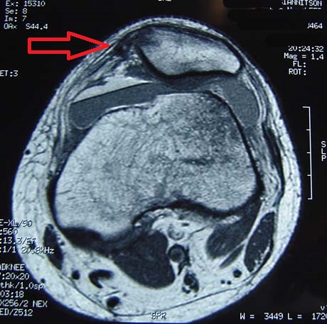
Medial patellofemoral ligament torn due to traumatic patella dislocation (red arrow)

CONSERVATIVE TREATMENT
THERAPY: 

CONSERVATIVE TREATMENT

Nonsurgical management of acute patellar dislocation has been the mainstay of treatment for this injury for centuries. Immobilization in a cylinder cast was recommended until more recently. So-called functional treatment has been recommended, with a reduction in recurrence and mobility. All nonoperative management options are associated with a high recurrence rate.

Goals:

Decrease pain and swelling
Limit range of motion and weight-bearing to protect healing tissues
Return muscle function
Avoid overaggressive therapy that may lead the patient into a patellofemoral pain
syndrome

Special functional braces for stabilization of patella.

OPERATIVE TREATMENT
THERAPY: 

OPERATIVE TREATMENT

Acute repair for young athletic patients who dislocated their patella via an indirect mechanism is generally recommended.
The MPFL ligament, which contributes greater than 50% of the resistance to lateral displacement of the patella in biomechanical testing, connects the medial border of the patella to the medial femoral epicondyle.
Most injuries of the medial patellofemoral ligament are avulsion from the distal femur. Blunt dissection is carried out, exposing the proximal half of the medial edge of the patella and the stump of remaining MPFL tissue. Nonabsorbable sutures are woven through the free edge of the avulsed MPFL using a locking stitch. In chronic cases where the injury was not treated in the first days after the injury, it is not possible to suture the ligament rupture. In these cases the reconstruction of a new autologous graft from the semitendinosus muscle is an appropriate and permanent solution to the problem.

REHABILITATION: 

Phase I for Immediate Postoperative – Weeks 1-6

Goals:

Control inflammation
Protect fixation
Activation of quadriceps and VMO
Full knee extension and minimize adverse effects of immobilization

Phase II – Weeks 6-8
Criteria for progression: Good quadriceps set, ~90 degrees of flexion, no signs of active
inflammation.

Goals:

Increase flexion
Avoid overstressing fixation
Control of quadriceps and VMO for proper patellar tracking

Phase III Week 8-4 months
Criteria for progression: No quadriceps extensor lag with SLR, nonantalgic gait, no evidence of
lateral patellar tracking or instability

Arthro Heal Clinic, www.arthrohealclinic.gr link image