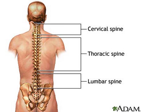
THE SPINE
THE CERVICAL SPINE
The seven bones in the neck are the cervical vertebrae. They support the head and connect it to the shoulders and body.
It is formed by first seven vertebra which are named as C1 to C7.
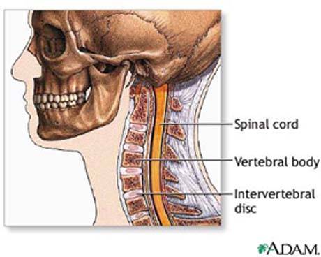
Anatomically, cervical spine starts where the top vertebra (C1) connects to the bottom of the skull. Normal cervical spine has a lordosis.That means it is curved with convexity on anterior aspect. It ends when C7 joins with first thoracic vertebra.
VERTEBRAE
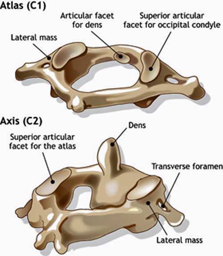
The base of the skull sits on top of C1.First vertebra or C1 is also called Atlas. It called so because it bears the direct weight of the skull, just as the mythical Greek hero Atlas bore the world on his shoulders.
Structure of atlas is unique.Two thickened bony arches form a large hole through the center of the atlas.
Because of its large size this opening is named as foramen magnum. Foramen magnum accommodates spinal cord which is wider here as compared to rest of it. The atlas has wide bony projections pointing out to each side.
The axis is the second of the seven cervical vertebrae, and is called such because it allows axial or rotational movement of the skull. The axis lies directly beneath the atlas vertebra.
They articulate at lateral articular surfaces and articulation of dens with atlas. Dens is a large bony knob on top of axis that points up and fits through a hole in the atlas.
The joints of the axis give the neck most of its ability to turn to the left and right.
C3-C6 vertebrae have a typical structures which is more or less same as discussed earlier in previous post. Following are special features/variations in these vertebrae.
The anatomy of the two upper vertebrae of cervical spine is totaly diferent. The structure of the ligament is extraordinary gives stability and secure.
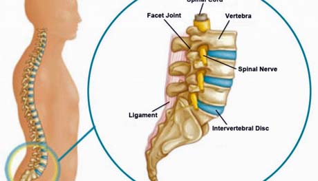
INTERVERTEBRAL DISC
The discs located in between each vertebrae function as shock absorbers and as joints.They are designed to absorb the stresses carried by the spine while allowing the vertebral bodies to move with respect to each other.
They made up of a strong outer ring of fibers called the annulus fibrosis, and a soft center called the nucleus pulposus.
The outer layer (annulus) helps keep the disc's inner layer intact. The annulus is made up of very strong fibers that connect each vertebrae together. The nucleus of the disc has a very high water content making it very moist.
SPINAL CORT AND NERVE ROOTS
The spinal cord extends from the base of the brain to the area between the bottom of your first lumbar vertebra and the top of your second lumbar vertebra. The spinal cord ends by dividing into individual nerves that travel out to your lower body and your legs.
This group of nerves at the end of the spinal cord is called the cauda equina, which is the Latin name for a horse's tail. For a short distance the nerve groups travel through the spinal canal before they exit out the neural foramen.
The dura mater is the protective membrane that covers the spinal cord. The dura mater forms a watertight sack around the spinal cord and nerves. The spinal cord is surrounded by spinal fluid inside this sack.
The nerves in each area of the spinal cord connect to specific parts of the body. The nerves of the cervical spine go to the upper chest and arms. The nerves also carry electrical signals back to the brain creating sensations.
Damage to the nerves, nerve roots, or spinal cord can lead to symptoms such as pain, tingling, numbness and weakness.
THORACIC SPINE
The section of the spine found in the upper back is called the thoracic spine. It goes from the base of the neck to the bottom of the rib cage. Knowing the main parts of the thoracic spine and how these parts work is important as you learn to care for your back problem.
The main function of the thoracic spine is to protect the organs of the chest by providing attachment for the rib cage. The 12 thoracic vertebrae are numbered T1 to T12. The range of motion in the thoracic spine is limited.
Viewed from the side, the cervical and lumbar regions have a lordotic, or slight concave curve, and the thoracic and sacral regions have a kyphotic, or gentle convex curve. The spine’s curves work like a coiled spring to absorb shock, maintain balance, and allow the full range of motion throughout the spinal column.
An abnormal curve of the thoracic spine is kyphosis, also called hunchback. Sometimes the spine abnormally curves from side-to-side, in a condition called scoliosis. A mild curvature (less than 20 degrees) is usually not noticeable or a health concern.
However, moderate curves (between 25 to 40 degrees) and major curves (over 45 degrees) are treated with braces or surgery. Scoliosis can put pressure on the heart and lungs as well as limit physical activity.
Normal spine from posterior and lateral view.
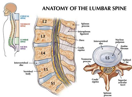
LUMBAR SPINE
The main function of the lumbar spine is to bear the weight of the body. The five lumbar vertebrae are numbered L1 to L5. These vertebrae are much larger in size for their weight-bearing function.
Lumbar spine (profile).
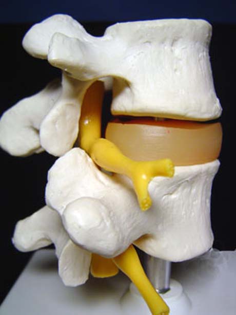
VERTEBRAL BODY, ARCH AND SPINAL CANAL
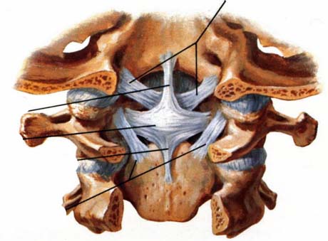
The body is the largest part of a vertebra, and is more or less cylindrical in shape.
On the back of each vertebra body are bony projections that form the vertebral arch.
The arch is made of two supporting pedicles and two arched laminae (Fig. 5).
The hollow spinal canal contains the spinal cord, fat, connective tissue, and blood supply of the cord. Under each pedicle, a pair of spinal nerves exits the spinal cord and pass through the intervertebral foramen to branch out to your body.
The facet joints of the spine allow back motion. Each vertebra has four facet joints, one pair that connects to the vertebra above (superior facets) and one pair that connects to the vertebra below (inferior facets)
INTERVERTEBRAL DISC
The discs located in between each vertebrae function as shock absorbers and as joints.
They are designed to absorb the stresses carried by the spine while allowing the vertebral bodies to move with respect to each other. They made up of a strong outer ring of fibers called the annulus fibrosis, and a soft center called the nucleus pulposus.
The outer layer (annulus) helps keep the disc's inner layer intact. The annulus is made up of very strong fibers that connect each vertebrae together. The nucleus of the disc has a very high water content making it very moist.
SPINAL CORD AND NERVE ROOTS
The spinal cord extends from the base of the brain to the area between the bottom of your first lumbar vertebra and the top of your second lumbar vertebra.
The spinal cord ends by dividing into individual nerves that travel out to your lower body and your legs. This group of nerves at the end of the spinal cord is called the cauda equina, which is the Latin name for a horse's tail. For a short distance the nerve groups travel through the spinal canal before they exit out the neural foramen.
The dura mater is the protective membrane that covers the spinal cord. The dura mater forms a watertight sack around the spinal cord and nerves. The spinal cord is surrounded by spinal fluid inside this sack.
The nerves in each area of the spinal cord connect to specific parts of the body. The nerves of the cervical spine go to the upper chest and arms. The nerves also carry electrical signals back to the brain creating sensations.
Damage to the nerves, nerve roots, or spinal cord can lead to symptoms such as pain, tingling, numbness and weakness.