CONGENITAL HIP DYSPLASIA
A CASE REPORT 1
Woman 44 years old with bilateral congenital hip dysplasia. The main symptom is severe and permanent pain at right hip joint 24 hours a day. The patient was not able to walk due to pain. She took amount of antiiflammatory drugs and pain killer for a long period. She has 3 daughters.
Congenital hip dysplasia bilaterally.
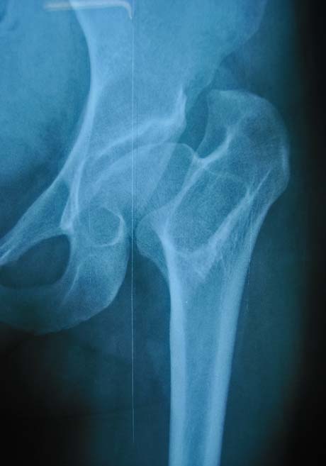
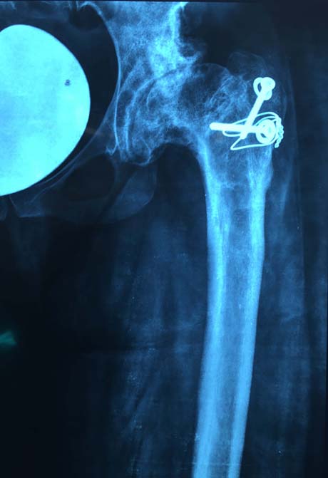
The head of the right femur has severe dysplasia and this is the reason for the hip osteoarthritis.
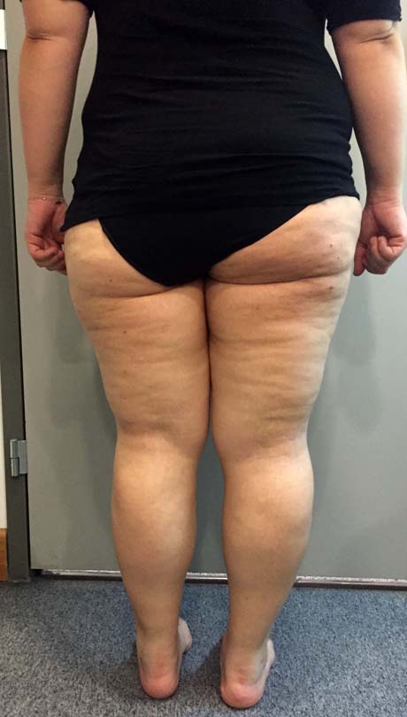
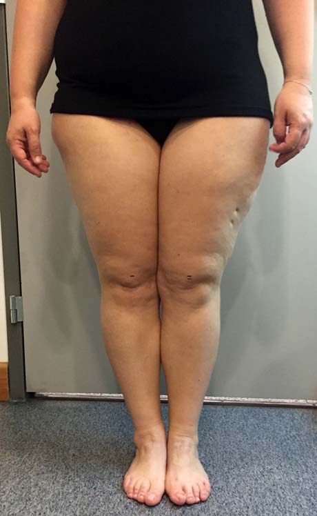
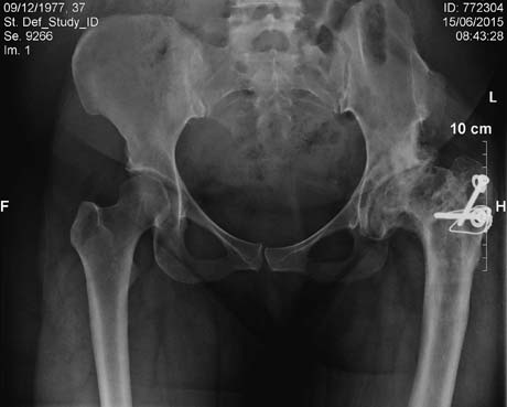
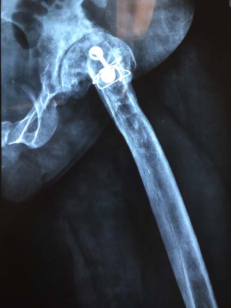
The right leg is shorter as compare as left 2,5 cm. Trochanteric tranfer when she was 16 years old (screw).
Left hip is dysplastic also but it is still painless.

Passive and active right hip flexion. The range of motion is not normal.

The high of the right femur is lower than left femur about 2,5 cm.
The heigh of the right leg is lower than left leg combine with the weakness of the pelvic muslce (hip abductors), that create limping.
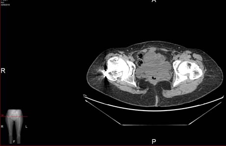
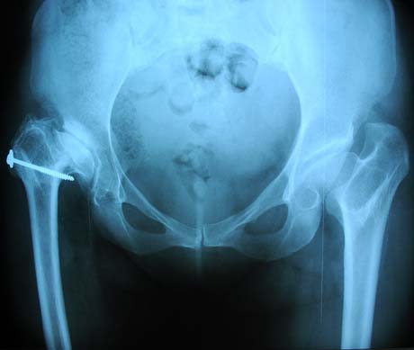
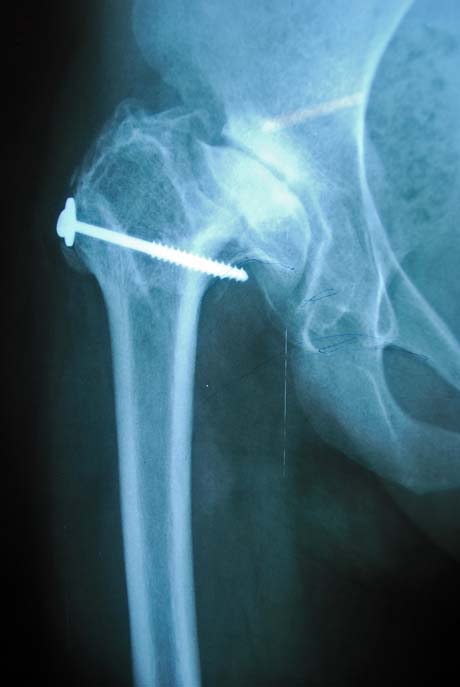
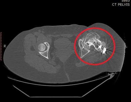
CT scan of the hip reveals the dysplastic hip bilaterally and severe osteoarthritis especially on right hip.
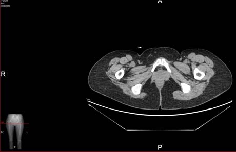
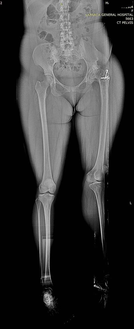
CT scan of the femurs reveal very narrow femur.
CT scan of the right hip joint reveals severe osteoarthritis that produce constant pain and this is the cause the patient cannot walk.

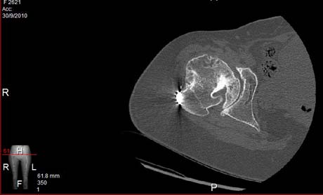
CT scan of the right femur reveals very narrow bone canal.
Cementless total hip replacement under general anesthesia.
The duration of the operation is 2 hours.
Two units blood tranfusion.
Next day physical therapy programm is started in bed
The 3rd day walking with assistive device without weight bearing under the observasion from physical therapist.
No weight bearing for 6 weeks.

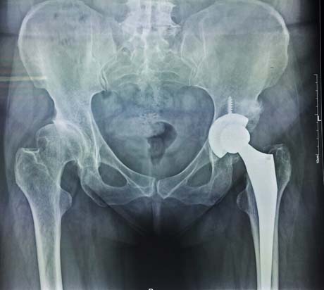
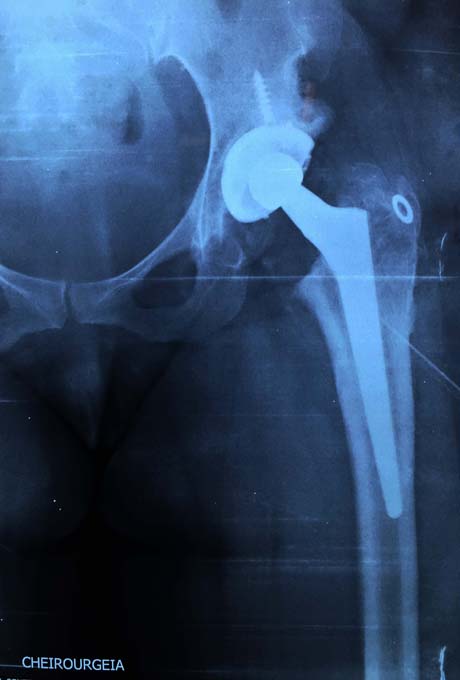
Cementless total hip replacement (right hip)

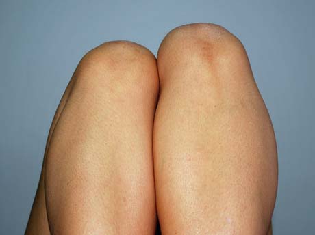
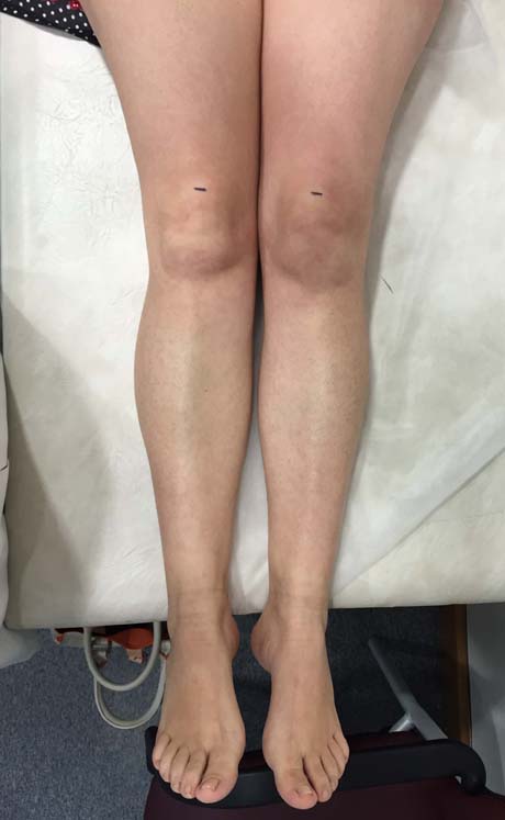
The heigh of the knee is equal post op
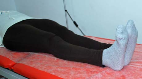
The leg length discrepancy is disappeared
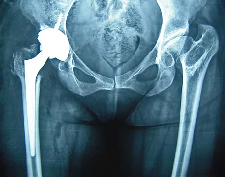
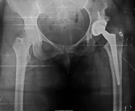
X-ray 8 years postop. The replacement does not show signs of wear. The clinical results are excellent
A CASE REPORT 2
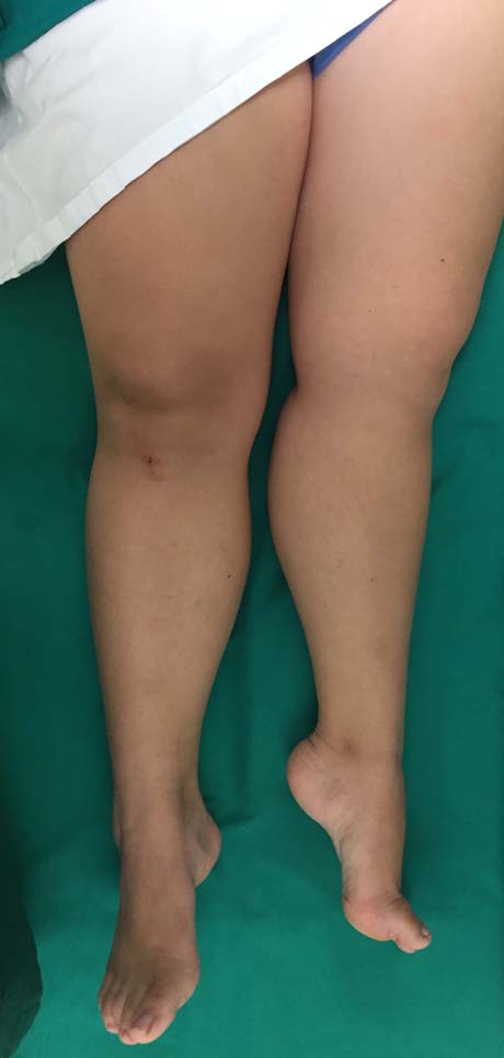
Female patient 44 years old suffers from severe arthritis in both hips of those due to developmental hip dysplasia - DDH. The left hip is in subluxation (upward displacement of the femoral head) due to congenital dysplasia of the acetabulum, so that the left leg is shorter and the patient has limping during walking.
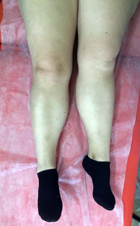
Leg length discrepancy (left lower limb is shorter 2.5 cm as compare to right)
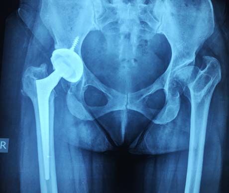
Pre op X-ray: severe hip osteoarthritis bilaterally. The left hip is subluxed due to developmental hip dysplasia DDH
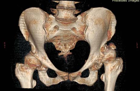
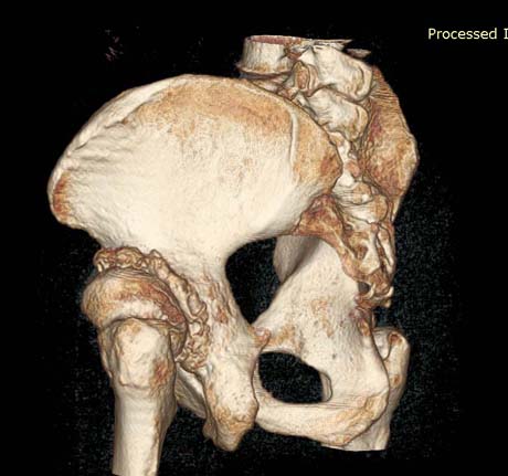
CT Scan - 3D reconstruction. Developmental hip dysplasia
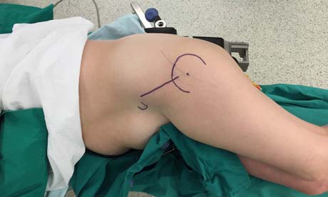
Pre op skin incision drawing (straight line)
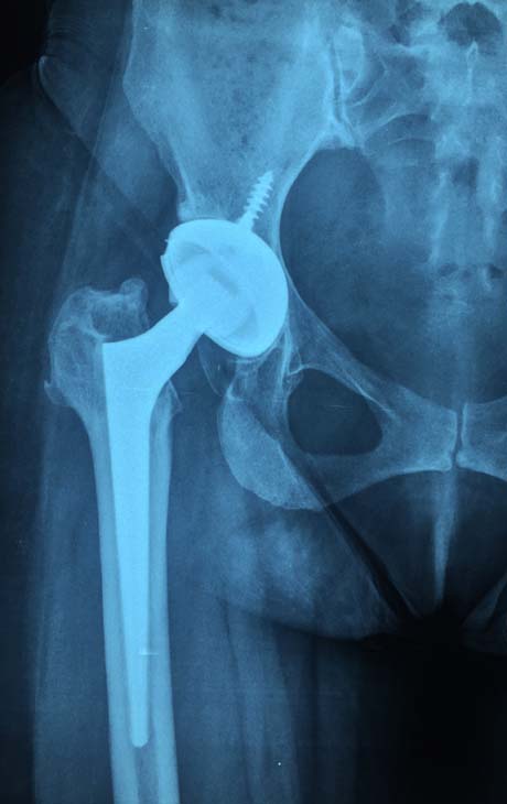
Post op X-ray. ALMIS hip arthroplasty left
The leg length discrepancy is restored
A CASE REPORT 3
Woman 35 years with congenital hip dysplasia left. In her medical history refers several operations which began in childhood and were designed to repair and improve the situation of the joint so as to become more
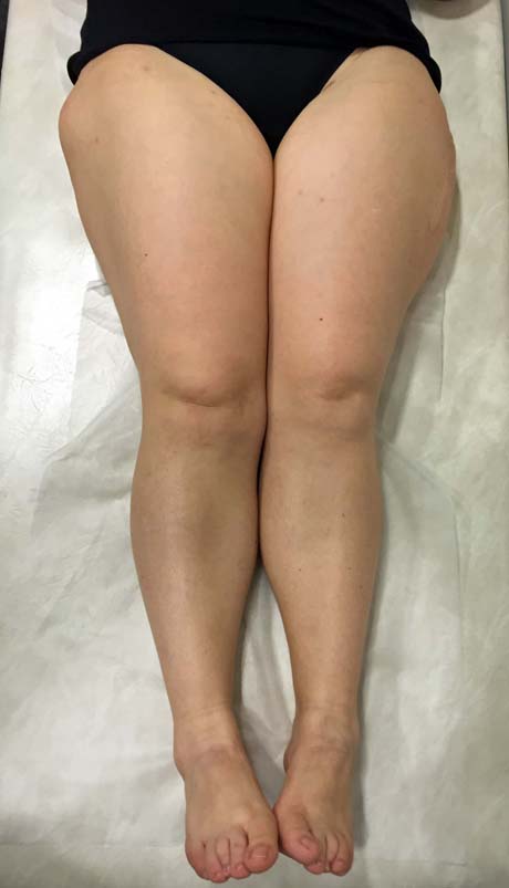
functional. The patient was examined by the writer and patient has severe limping of the left leg and leg length discrepancy 5 cm and distortion - external rotation of the left lower limb, combination which makes walking very difficult and laborious.


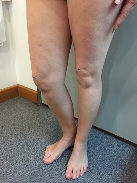
Prior to surgery. Patient woman 35 years with congenital dysplasia of the left hip. Leg length discrepancy and distortion in external rotation of the left leg is particularly evident
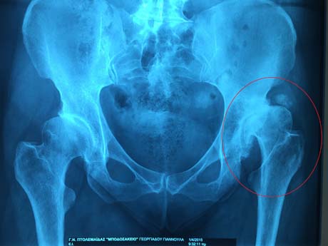
Pre op X-ray, hip arthritis due to developmental left hip dysplasia
CT-scan severe hip osteoarthritis left (circle)
CT-scanogram
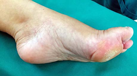
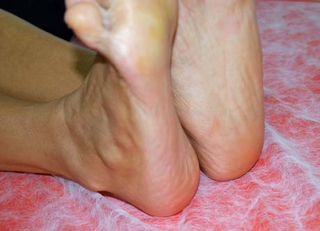
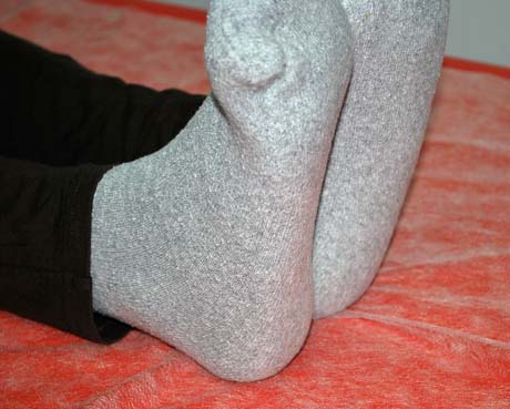
Burden on metatarsal heads due to acquired deformity of the foot to the compensatory Leg length discrepancy (equinus)
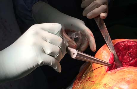
Post op X-ray, total hip arthroplasty of the left hip
Four months post-op, the mechanical axis of the left lower limb is completely restored