CARTILAGE LESIONS OF THE TALUS

OSTEOCHONDRAL LESION OF THE TALUS

OSTEOCHONDRITIS DISSECANS OF THE TALUS

Large osteochondral defects of the talar dome, typically involving the talar shoulder (transition of superior dome cartilage to the medial or lateral cartilage), and also often associated with large-volume subchondral cysts.
Cartilage lesions of the talus are focal injuries of the articular cartilage and subjacent subchondral bone may also be damaged.

These osteochondral defect primarily occur as a result of acute and chronic ankle sprains, of repeated microtrauma resulting from chronic instability or fo axial malalignment of the hindfoot.


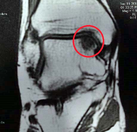
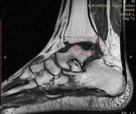
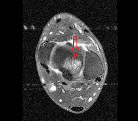
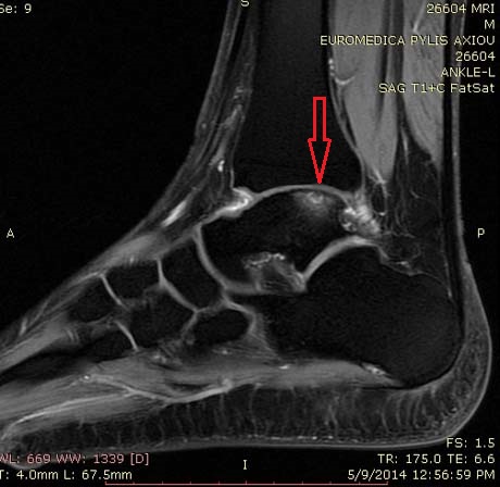
MRI, chondral injury of the medial talus


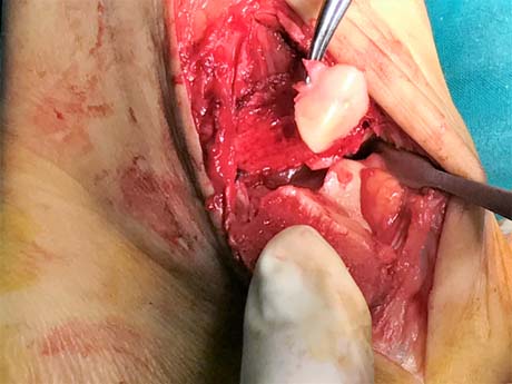
Part of ankle joint cartilage from the upper medial articular surface which is fully detached because the osteochondritis dissecans


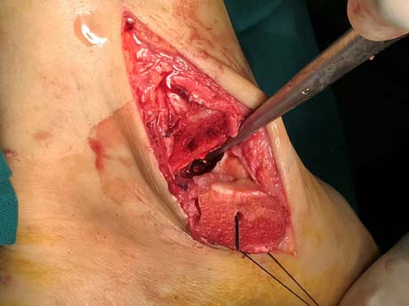
The bone defect created in the upper and medial articular surface of the talus after removal of the detached portion

Anatomy of an osteochondral lesion —three zones can be distinguished

Cartilage: Compared to the knee joint, the ankle cartilage is 1–1.5 mm thinner and less elastic, and has chondrocytes that are more resistant to osteoarthritic damage.

Subchondral layer: The subchondral bone is essential for the stability of the cartilage. It fixes the cartilage stably to the bone.

Bone: Bone provides nutrition to the cartilage. Changes of the cancellous structure leading to cysts and sclerosis can be found.
 

PATHOGENESIS

The pathogenesis for osteochondral lesions of the talus(OLTs) is not fully understood.
Theories include:

• Trauma
• Idiopathic focal avascular necrosis
 

SYMPTOMS

Persistent load-dependent pain in the ankle joint severely restricts patients in their work and in their leisure activities.
Patients may or may not report a history of trauma
Ankle pain, typically involve the anterior aspect of the ankle, is a common complain.


DIAGNOSTIC STUDIES

• Plain radiographs
• MRI (Magnetic Resonance Imaging)
• CT-scan


CONSERVATIVE TREATMENT
THERAPY: 

NONOPERATIVE MANAGEMENT

Non-surgical
treatment involves a combination of rest or restriction from
strenuous activity, an initial period of non-weight-bearing
with special brace immobilisation, and subsequently protected weight
bearing with gradual mobilisation thereafter.
 

  • Activity modification
  • Bracing
  • Physical therapy if associated ankle instability
  • Nonsteroidal anti-inflammatories or COX-2 inhibitors
  • Corticosteroid injection
  • Viscosupplementation
  • PRP (Platelet Reach Plasma)

The new orizon in chondral damage therapy is intrarticular injection of mesenchymal stem cells

OPERATIVE TREATMENT
THERAPY: 

SURGICAL TREATMENT

Surgical treatment is considered with failed conservative management. 

Surgical management strategies for osteochondral lesions of the talus include excision with or without techniques forstimulation of fibrocartilage growth such as microfracture, curettage, abrasion, or transarticular drilling.

If the fragment is large enough, it may be secured to the talar dome through
retrograde drilling, bone grafting, or internal fixation.

Other surgical strategies include cancellous bone grafting and osteochondral transplantation through osteochondral
autografts, allografts, or cell cultures.
 

ANKLE ARTRHOSCOPY

MICROFRACTURE

Microfracture is indicated as first-line treatment for focal Outerbridge grade III and IV lesions under 3–4 cm2 in an otherwise nonarthritic knee in a symptomatic, active, and relatively young patient willing to respect the postoperative rehabilitation requirements.  The microfracture techniques is originaly an arthroscopic procedure.


Posterior arthroscopic portals


Ankle arthroscopy, anterior.

Ankle joint arthroscopy, posterior

Special instrument for microfracture and nanofracture technique

For older and low-demand patients, microfracture may be useful even in lesions larger than 3–4 cm2.
The functional principle of microfracturing is based on the release of multipotent mesenchymal progenitor cells, cytokines and growth factors from the subchondral bone.

POSTOPERATIVE
Cold therapy is typically used for 1 to 7 days postoperatively.
Crutch-assisted touchdown weight-bearing ambulation (10% of body weight initially) is prescribed for 6 to 8 weeks, depending on the size of the lesion.
For most patients, 6 to 8 weeks is adequate time to limit weight bearing. However, for patients with small lesions (<1 cm diameter), weight bearing may be initiated earlier.
 

AUTOLOGOUS MATRIX INDUCED CHONDROGENESIS AMIC  

is an innovative biological surgical procedure developed for the treatment of traumatic chondral or osteochondral lesion. This unique single – step procedure combines the microfracture method with the application of Chondro – Glide, a porcine collagen type I/III matrix.

The super clot formed as a result of haemorrhage due to microfracture. This clot stabilised by Chondro-Gide matrix and fibrin glue.

This scaffold is suitable that enchance the chondrogenic differentiation of mesenchymal stem cells and, in combination with fibrin glue stimulates chondrocytes to enhance proteoglycan deposition.


The collagen membrane covers the chondral defect

AUTOLOGOUS CHONDROCYTE IMPLANTATION

Injuries to joint surfaces can result from acute high-impact or repetitive shear and torsional loads to the superficial zone of the articular cartilage architecture. The use of autologous chondrocyte implantation is promising and is associated with several potential long-term benefit.

Autologous chondrocyte implantation is ideally suited for symptomatic deep chondral lesion along the talus dome region. High-demand patient between 15-55 years of age with excellent motivation and potential for compliance are the best candidates.

However autologous chondrocyte implantation is a viable option for a symptomatic patient with a lesion of >2 sq.cm but <12 sq.cm. and for a patient who continues to have pain after mosaicplasty or microfracture procedure.

Bone involvement is not contraindication, but staged or concomitant autologous bone grafting should be undertaken when the bone involvement is deeper than 6-8 mm.

Autologous chondrocyte transplantation is not a treatment for osteoarthritis (general joint disease), gout, and rheumatoid arthritis or other systemic joint diseases.

Osteochondral lesion of the talus (red arrow)

Autologous chondrocyte are arthroscopicaly harvested from a less weight-bearing area, commercially extracted from the harvested cartilage (200-500mg), and multiplied in vitro (labor cell culture).
Elective reinplantation is performed 4-6 weeks after cartilage harnesting by debridement of the defect to an intact margin, carelully avoiding osseous bleeding from the bed of the defect.

Cultured chondrocyte ready for implantation


Special shape analogue of the lesion shape.

POSTOPERATIVE

Postoperatively protected weight-bearing are restricted for 6 weeks. The crutches is necessary for walking.
The physical therapy program started 2nd post-op day with passive movement until 4th week.
Therapy concentrates on quadriceps activation and isometric straight leg raises. After the 6th week the patient can walk without aid devices with full weight-bearing. Full weight bearing and full range of motion are achieved. Closed-chain exercises and functional training are started.

After 12th week Strengthening and functional training are progressed. Return to full activities is delayed for at least 8 months to protect the lesion as it matures.

Return to participation in pivoting sports is usually allowed by 12 months.

REHABILITATION: 

MICROFRACTURE TECHNIQUE

Cold therapy is typically used for 1 to 7 days postoperatively.

Crutch-assisted touchdown weight-bearing ambulation (10% of body weight initially) is prescribed for 6 to 8 weeks, depending on the size of the lesion.

For most patients, 6 to 8 weeks is adequate time to limit weight bearing. However, for patients with small lesions (<1 cm diameter), weight bearing may be initiated earlier.


ΑUTOLOGOUS CHONDROCYTE IMPLANTATION

Postoperatively protected weight-bearing are restricted for 6 weeks. The crutches is necessary for walking.

The physical therapy program started 2nd post-op day with passive movement until 4th week.

Therapy concentrates on quadriceps activation and isometric straight leg raises. After the 6th week the patient can walk without aid devices with full weight-bearing. Full weight bearing and full range of motion are achieved. Closed-chain exercises and functional training are started.

After 12th week Strengthening and functional training are progressed. Return to full activities is delayed for at least 8 months to protect the lesion as it matures.

Return to participation in pivoting sports is usually allowed by 12 months.

Arthro Heal Clinic, www.arthrohealclinic.gr link image