ANTERIOR KNEE PAIN
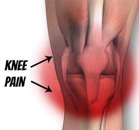
The pain in the anterior surface of the knee is a common clinical entity that relates to athletes - jumper's knee, runner's knee - but also to people who are not engaged in a sport regularly or amateur.
The pain of knee referred internationally as' anterior knee pain'- or 'patellofemoral pain syndrome' and the therapeutic approach should be multifactorial and including the involvement of the patient himself.
The patient reports mainly pain and stiffness in the knee prolonged immobility of joints (prolonged sitting position), or during activities that require bending of the knee such as raising and lowering the ladder, deep kneeling.
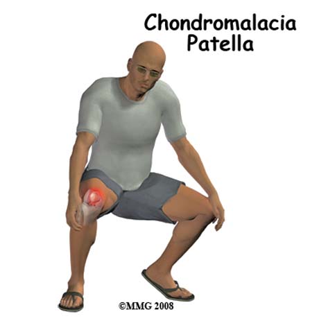
Pain during effecting deep seating knees -squating
The cause of patellofemoral pain is multifactorial implicated factors such as improper alignment of the patella, disturbances in the mechanical axis of the lower end (valgus knee, external rotation of the tibia, etc.), as well as sustained and persistent stress of said hinge sports or professional activities.
CAUSE
OVERUSE SYNDROME
In many cases, patellofemoral pain syndrome is due to prolonged and difficult physical activity which causes a particular stress in the joint such as jogging, lifting and lowering the ladder. It can also be due to a sudden change of physical activity such as starting a sport after prolonged abstinence from sports. It can also be due to variation in the frequency of physical activity or duration of fulfilling the example increased kilometers traveled by a runner. The sudden weight gain extra burden joint.
PATELLAR MALALIGNMENT
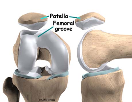
The kneecap (patella), 'nests and smoothly slide within the femoral trochlea (femoral or trochlear groove), during flexion and extension of the knee
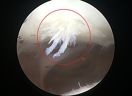
Arthroscopical view: Patellar articular cartilage damage due to chronic patellofemoral impingment syndrome
Radiographic view 'sunrise view', normal imaging of patella in the femoral trochlea
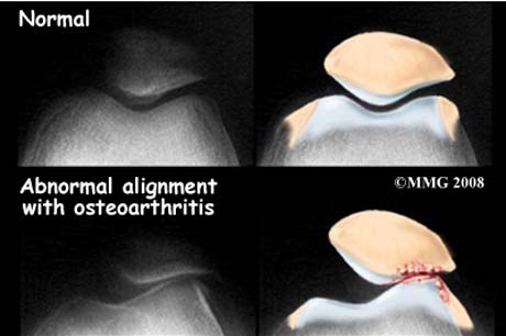
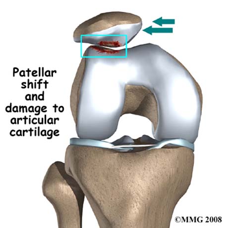
The forces exerted on the patella sometimes tend to shift from its normal position thereby causing damage of the articular cartilage which eventually causes pain.
The arrow shows the point of increased patella pressure on the outer surface of the femur. The patient feels intense knee pain especially when climbing and falling down stairs due to local development of arthritis.
Severe patellofemoral osteoarthritis
The anterior knee pain may however be due to abnormal slipping patella in substrate the femoral trochlea. In this situation the patella is pushed away from its normal position during knee flexion. This causes increased pressure and wear between the articular surfaces of the patella and femoral trochlea stimulating round synovial bursa.
FACTORS THAT CAUSE ABNORMAL SLIPPING PATELLA INCLUDE:
ANATOMIC FACTORS
1. The femoral trochlea, the substrate i.e. sliding patella is shallow and may not include the patella during flexion and extension motion. This results in the extrusion of the patella to the outside and simultaneously creating an increased pressure conditions between the articular cartilage and premature wear.
2. The high position of the patella -patella alta- with respect to the normal position creates conditions abnormal friction between the two bone pain resulting appearance.
3. valgus knee because the knee extrude outwardly
4. Women because of the wider pelvis than men tend to have problems with their knee pain of adolescence.
From the pelvis starts the quadriceps, the main tensile muscles of the knee and the vector that traction on the patella pushes outwards.
5. The outward shift tibias - externally rotated tibia - people that walk with their open end feet, creating traction conditions out of patellar pain resulting appearance.
6. Stiff patella due to tight ligament structures around the joint.
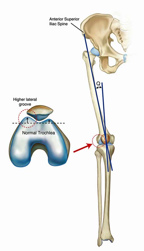
The Q angle shows the force exerted by the quadriceps on the patella. The greater the Q angle both quadriceps tends to 'pull' the patella outwardly pulling it from the completely normal anatomical position. Women said open basin with respect to the man exhibit greater Q angle and hence greater sensitivity patellofemoral articulation
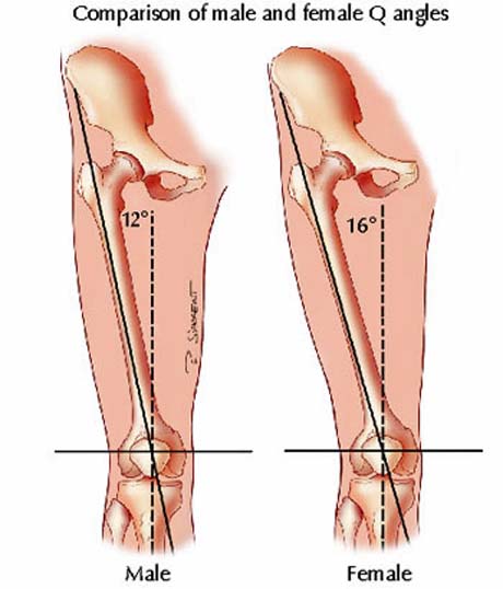
Comparison Q angle with respect to gender. Man (male), woman (female)

Female Male
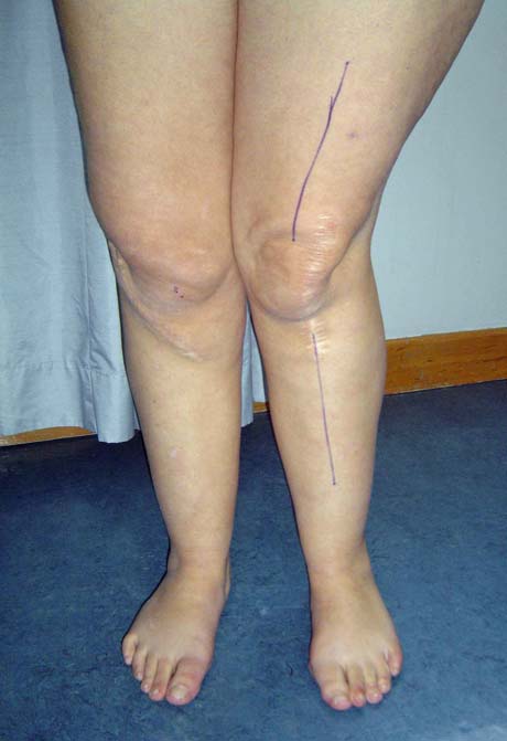
Failure of the mechanical axis of the left lower limb due to old injuries creates conditions subluxation of the left patella and pain in the joint
Genu recurvatum causes severe disturbance in the sliding of the patella and pain in the joint
MUSCLE FACTOR
The weakness of the quadriceps muscle is one of the most common causes of anterior knee pain. The quadriceps muscles helps the patella is maintained in the femoral trochlea creating painless joint motion conditions. The quadriceps muscles may lose much of its strength rapidly after an injury to the lower end, and therefore require a special reinforcement rehabilitation program to quickly return to its normal state.
'Tight hamstrings muscles'. The hamstring muscles inserts in the tibia and if it is inflexible through many times of great muscular power, creating increased pressure of the patella on the substrate resulting in severe pain.
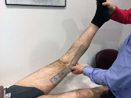
CLINICAL EXAMINATION X-Ray CT, MRI
Normal patellar tracking in young male
Patellar maltracking produce anterior knee pain and chondromalacia in adolescent female 14 years old
Abnormal course of the patella during flexion and extension of the knee - bounce - because of abnormal anatomy in the patellofemoral joint in girl 12 years
Boy 8 years old with hemophilia. Disorder slipping patella in both knees.
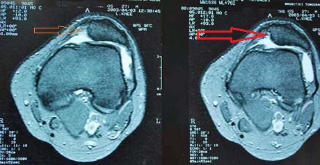
Woman 23 years old with patellar maltracking due to trochlea dysplasia. Patellar subluxation in every movement flexion and extension of the knee
The examination made by the Orthopaedic surgeon aims to clarify the cause of pain and not just suppress the pain with painkillers and anti-inflammatory drugs. The investigation and identification of the cause is the cornerstone of treatment of the problem.
This may have clinical evaluation of the knee more than once and conducting diagnostic work like simple radiological examinations MRI, CT scan, or CT scanogram for detailed imaging of bone and muscle joint structures and mechanical axis of the lower limb .
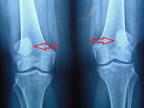
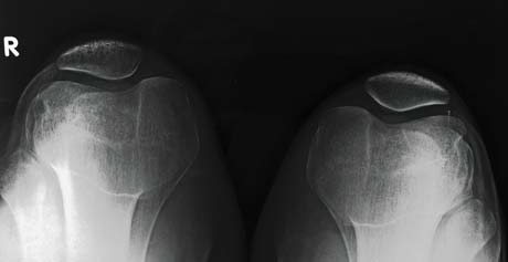
X-Ray knee bilaterally. The patellae are outside of their normal positions (upper and out of normal)
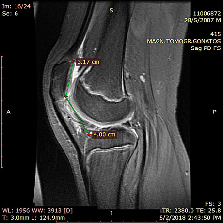
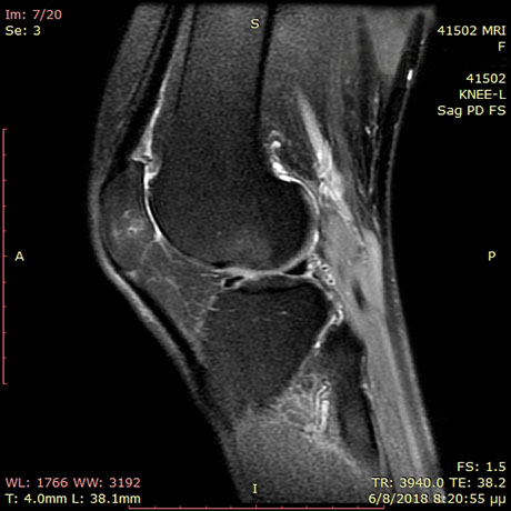
Knee Magnetic resonance imaging MRI- sagittal section in adolescent. The elevated position of the patella (patella alta), causes significant damage to the articular cartilage thus causing pain.
The same patient. The position of the patella related with the trochlea is not normal and that can produce anterior knee pain and early arthritis.
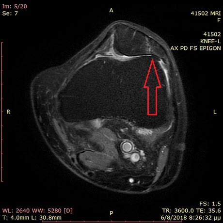
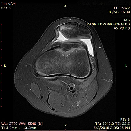
Magnetic resonance imaging MRI, of the left knee showing the outward patella subluxation due to dysplasia of the femoral trochlea
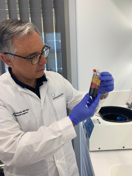
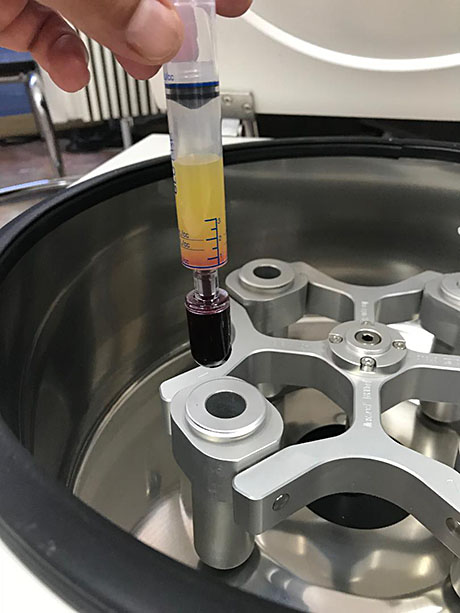
PRP - GROWTH FACTORS PLATELET RICH PLASMA
PRP - PLATELET REACH PLASMA