
KNEE HEMANGIOMA IN GIRL
A CASE REPORT
Girl, five years old suffering from swelling in his left knee and pain. The symptoms began at the age of 3.5 years. The patient experienced occasional severe edema (swelling) in the left knee with inflammatory signs such as increased redness and increased local temperature of the affected joint. Limitation of knee flexion during the final degrees accompanied by intense pain. The first diagnosis was juvenile rheumatoid arthritis was until the age of 5 where the magnetic resonance imaging of the knee showed the lesion. During the clinical examination was palpable mobile mass without causing severe pain to the child.
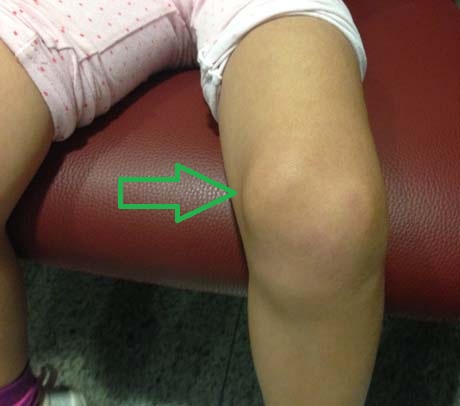
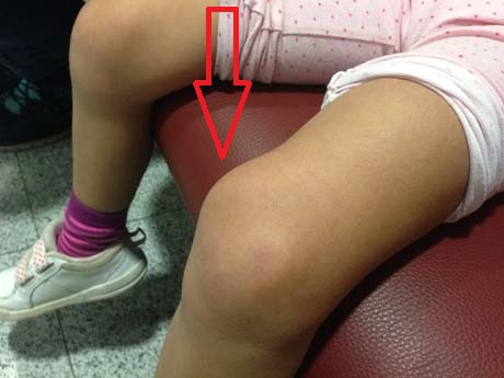
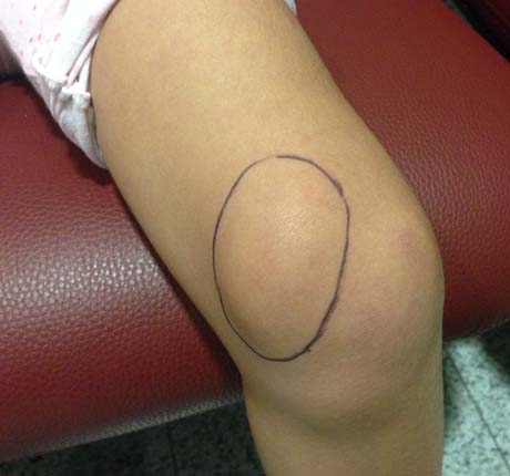
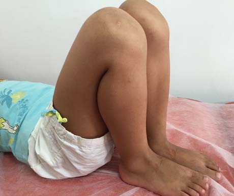
Girl 5 years old with a palpable mass in the left knee medial up side (arrow)
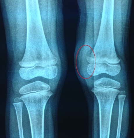
The radiological examination of the knees shows soft tissue mass in the medial side of the left knee (circle)
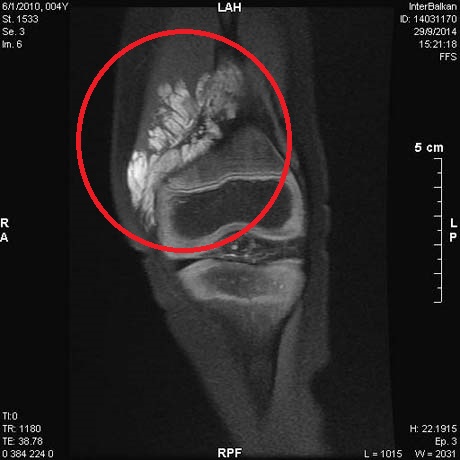
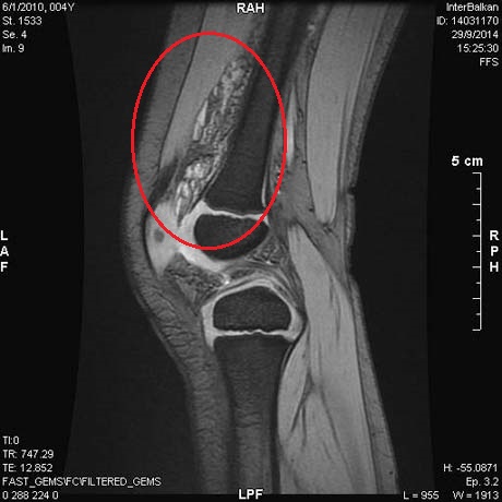
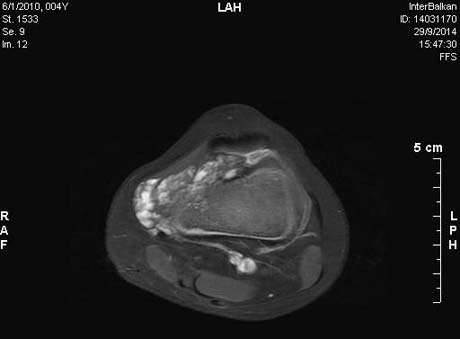
MRI left knee. Emergence lobed mass in the inner and the upper region of the joint
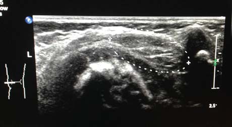
Ultrasonography knee. Knee lobed mass
After Investigations and clinical study of the small patient, the decision was arthroscopy of the left knee joint to make the evaluation of the extension of the mass and any damage is caused to the articular cartilage or other knee tissue. Then we performed open surgery for the evaluation and better control of major vascular mass and of course the final exclusion of this, safely.
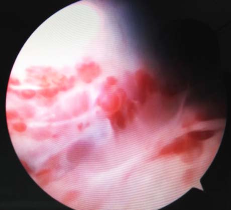
Arthroscopic image of the mass. This image is typical hemangiomas mass
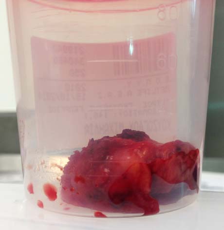
The hemangiomas mass after removal. The size is significantly smaller because the blood has been removed from within
The mass was sent for histological examination where diagnosed hemangioma of the knee. The patient did not receive any form of adjuvant therapy after surgery.
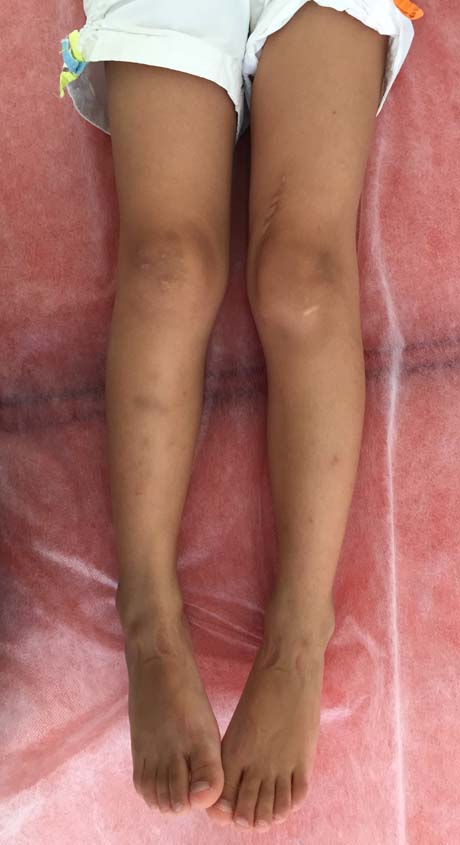
Four months post-op
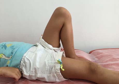
The mobility of the joint is fully restored without pain
The monitoring of small patient continued for two years after surgery. The girl showed no recurrence of the disease. The joint motion range was returned to 100% and the pain disappeared for all.
The inflammatory signs of the knee joint as swelling, warming and redness ever disappeared after surgery. The initial diagnosis was as juvenile rheumatoid arthritis was incorrect and could result in error therapeutic roads medication which would have no effect on hemangioma.
Hemangioma is a benign tumor derived from the vascular tissue. After successful surgical removal of relapse rates are extremely low. In large hemangiomas that nutrient arterial strain is large there may be a need embolism initially and after surgery excused.